You are incorrect - our patient's electrocardiogram is entirely normal.
Click on the links to learn about this ECG:
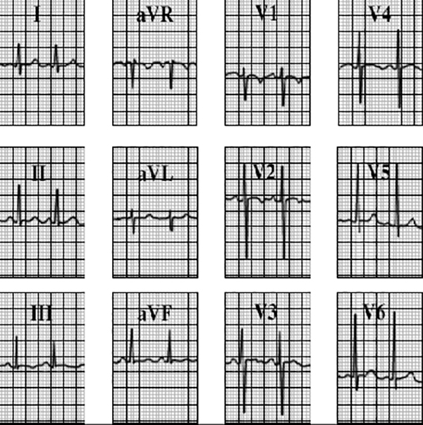
Your choice: Persistent juvenile pattern
The classic juvenile ECG pattern shown here demonstrates inverted T waves in leads V2 and V3 with some slight T wave inversion in lead V4.
This pattern is typically seen in normal young children, but may persist past adolescence in some normal patients.